
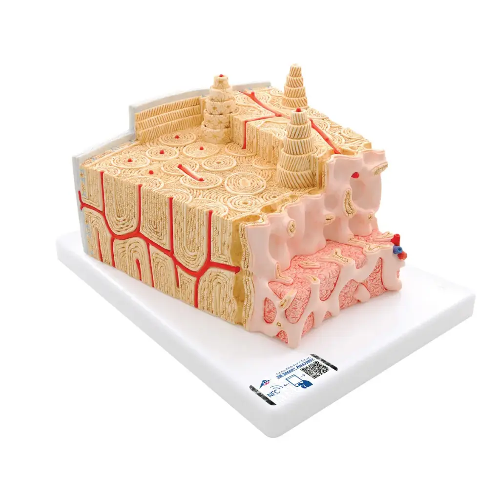





Bone structure model
| Brand: 3B |

Référence |
1000154 |
Features
- Microanatomical model of lamellar bone representing a detailed three-dimensional section of bone structure.
- 80x magnification for precise observation of the various constituent elements of bone.
- Representation of a tubular bone with transverse and longitudinal sections at different anatomical levels.
- Section of bone marrow with visualization of its internal structure on two planes.
- Osteons (Havers systems) clearly identifiable to understand the organization of compact bone.
- Havers and Volkmann canals illustrating the vascular and bone communication networks.
- Compact and spongy substance represented to compare their roles and characteristics.
- Visualization of the endosteum and cortical substance in their anatomical context.
- Presentation of osteocytes and their arrangement within bone tissue.
- Advanced educational resource designed for teaching bone anatomy, histology and physiology.
- Mounted on base for stable presentation in classroom or laboratory.
- Digital twin included accessible on smartphone, tablet and computer.
- Interactive rotation and zoom enabling detailed exploration of the digital model.
- Interactive anatomical landmarks with display of corresponding structures.
- Augmented Reality (AR) function to visualize the model in a real environment.
- Integrated anatomy quiz with instant results and knowledge assessment.
- Annotation and drawing function with saving and sharing of personalized images.
- Digital note-taking facilitating learning and memorization.
- Online and offline access to 3D anatomical content.
- Available in 11 languages for international use.
"An exceptional tool for visually understanding the complex architecture of bone and making bone anatomy immediately accessible."
| Technical Features | Details |
|---|---|
| Product Type | Microanatomical model of lamellar bone |
| Manufacturer | 3B Scientific® |
| Structure Represented | Three-dimensional section of a lamellar bone |
| Magnification | 80x |
| Type of Bone Illustrated | Tubular bone |
| Anatomical Sections | Transverse and longitudinal sections at different levels |
| Bone Marrow | Internal structure represented on two cutting planes |
| Visible Anatomical Structures | Osteons (Havers systems), Havers canals, Volkmann canals, osteocytes, endosteum, cortical substance, compact substance and spongy substance |
| Support | Supplied mounted on base |
| Primary Use | Teaching bone anatomy, histology and bone structure |
| Digital Twin Included | Yes |
| Digital Compatibility | Smartphone, tablet and computer |
| Digital Functions | 3D rotation, zoom, interactive anatomical landmarks, augmented reality, anatomy quiz, annotations, note-taking |
| Content Access | Online and offline |
| Available Languages | 11 languages |
What is this microanatomical bone model for?
This model is designed to illustrate in detail the internal structure of a lamellar bone and facilitate learning of bone anatomy and histology.
What is the magnification level of the model?
The model represents bone structure with 80x magnification to make essential microscopic elements visible.
Are osteons visible on this model?
Yes, osteons, also called Havers systems, are clearly represented and easily identifiable.
Which anatomical structures are represented?
The model shows in particular osteocytes, Havers canals, Volkmann canals, endosteum, compact substance, spongy substance and cortical substance.
Is bone marrow represented?
Yes, the model includes a section of the internal structure of bone marrow observed on two cutting planes.
Is this model suitable for teaching?
Yes, it is particularly suited for educational institutions, laboratories and health science training.
What digital features are included?
The digital twin allows model rotation, zoom, display of anatomical landmarks, augmented reality, quizzes, annotations and note-taking.
On which devices is the digital content accessible?
Digital content is accessible on smartphone, tablet and computer, with possible use online and offline.
In how many languages is the digital content available?
Digital anatomical content is available in 11 languages.